Primarily, the microscope detects minute changes in refractive index (RI) throughout the specimen volume by mesuring phase changes affecting light travelling in different directions. It combines principles of phase microscopy with 3D holographic reconstruction and deconvolution, achieving lateral resolution of 200 nm. As RI is generally proportional to density this method is particularly suitable for imaging and tracking of dense cytoplasmic bodies (stress granules, P-bodies) or sub-nuclear domains (e.g. PML, Cajal bodies, nucleolus). Hence it is an excellent tool for monitoring and tracking formation of membrane-less organelles that are formed by liquid-liquid phase separation, often with the help of intrinsically disordered proteins and RNA.
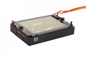
Our setup is equipped with epifluorescence module (2D only) so it also allows detection and co-localization of genetically encoded protein markers and nucleic acids (DAPI). We have also stage top incubator (okolab) with temperature, gas and humidity control that allows long-term (days) live cell imaging.


Below are the actual parameters of the inverted microscope installed here:
- Microscope Objective: Air 60x magnification with low power laser (λ = 520nm, sample exposure 0.2mW/mm2)
- Resolution: Δxy = 200nm; Δz = 400nm
- Field-of-View: 85 × 85 × 30 μm
- Depth-of-Field: ~ 30µm
- Tomography Frame Rate: 3D image rate 0.5fps, full self-adjustment
- Accessible sample stage: 60 mm of free access to the sample stage for sample manipulation
- up to three pre-configured epifluorescence (2D) channels: blue (DAPI), green (fluorescein, GFP), red (TritC, mCherry)
- works best with 35 mm imaging grade (glass bottom cover slip) petri dish e.g. from ibidi

- Data collection can be programmed to interlace RI holographic images with fluorescence for low dose, non-bleaching time-lapse imaging.
The system is located in BSL2 lab next to cell culture to allow work with live cells and potentially infectious agents.
If interested in using the system or getting trained please contact Roman Tuma: rtuma(at)prf.jcu.cz or Zdenek Franta zfranta(at)prf.jcu.cz
The system is easy to set up and brief demo and/or training session can be arranged upon request (e.g. alongside our own imaging session)